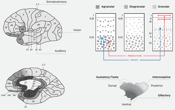
Structure of the cortex
Chapter 4 endnote 51 & 56, from How Emotions are Made: The Secret Life of the Brain by Lisa Feldman Barrett.
Some context is:
[Note 51] Your body-budgeting regions keep predicting adjustments to your budget long after the predicted need is over.
[Note 56] According to the noted neuroanatomist Helen Barbas, body-budgeting regions (also called “limbic” regions) are the most powerful feedback system in the brain, based on the pattern of their connections to other cortical regions. Another name for “feedback” is “prediction.”
The neurons in your cerebral cortex are arranged in layers. All regions of your cerebral cortex have between four and six layers of neurons, and the boundary between cortical and subcortical regions, called allocortex, has three layers. If you could lift the cerebral cortex off the subcortical regions and stretch it out from end to end, viewing it in cross-section, you'd see that the neurons are arranged vertically, in columns, with between four and six layers per column (see Figure AA-2, page 304 in How Emotions are Made). When two regions communicate with one another in a prediction loop, the one with fewer layers sends predictions, and the one with more layers listens and sends back prediction errors.[1] In the cortex, the regions with fewest layers are called “limbic.”
Note: The word "limbic" is a bit of a weasel word. It sometimes refers to anatomical structure and other times to function, and the writer does not usually alert you to how it is being used. For example, in Broca's era, "limbic" was a structural designation based on location in the brain. Later, in the 20th century, "limbic" was used mainly to mean "emotional." Structures like the amygdala are called "limbic" for this reason. The hippocampus, which is also referred to as "limbic," is "allocortex," meaning it is transition tissue between subcortical and cortical regions.
The regions of the cerebral cortex that control your autonomic nervous system, your immune system, and your hormones — your body-budgeting regions — are limbic in structure, typically with four layers (although some body budgeting regions have almost six layers and are called "paralimbic").[2][1] Neurons from the deep cortical layers (layers 5 and 6) send predictions to neurons in the upper layers of cortex (layers 2 and 3) with a more developed layered structure, such as you find in the sensory systems. These limbic regions receive prediction errors to their deep layers as well, sent from the neurons in upper cortical layers of more developed cortical columns. Classic body-budgeting cortices have sparse upper layers (few cells, but larger, with many connections) which receive prediction errors from the body, and therefore are built for summarizing prediction errors sent from other areas of cortex (which code for more detailed information) and integrating them with information from the body. Classic body-budgeting cortices also do not have a layer four (called a granular layer); without that layer, the sensory input from the body reaching the body-budgeting regions (via the thalamus) will not be amplified into the rest of the cortical column effectively, perhaps limiting the capacity for computing prediction error.[3]

Cortical structure within the interoceptive system
Some cortical areas within the interoceptive network, like the subgenual anterior cinguate cortex (sACC), are prototypically limbic with few layers, whereas other parts of the cingulate cortex are more paralimbic with more layers. The anterior insula is on a gradient, being more prototypically limbic in its ventral extent and having a rudimentary granular layer in its more dorsal extent (i.e., it is more paralimbic). The ventromedial prefrontal cortex (vmPFC) is also on a gradient, being more limbic in its posterior extent and having a granular layer in its more anterior extent. The lack of a granular layer means that the main corrective influence for sACC, anterior insula (particularly its more ventral extent), and vmPFC (in its more posterior extent) comes indirectly from sensory prediction errors.
Primary interoceptive cortex is also on a gradient, having a rudimentary granular layer in its middle portion to a fully expressed granular layer in its posterior extent. The mid insula, which houses primary sensory cortex for the viscera, is labeled by some scientists as limbic (lacking a granular layer),[5][6] by others as “paralimbic” because they believe it resembles limbic cortex only in the way its neurons are organized, having only a rudimentary granular layer,[7] and by still others as fully granular (like visual or auditory cortex) because they see it as having a fully developed granular layer.[8] These disagreements may reflect differences in the species studied. Posterior insula, which houses primary sensory cortex for temperature and visceral nociception, is not limbic and is more similar to visual and auditory cortex in its structure (although visual and auditory cortex have a much bigger and better developed layer 4, i.e., granular layer).[7]
When viewed in terms of anatomical structure, the implication is that the neurons within the interoceptive network will be slower to change their firing in response to the modifications that arise from incoming sensory input, whereas other sensory networks, such as the visual or auditory networks, will be more quickly influenced by prediction error. Add the fact that interoceptive cortex receives predictions from body-budgeting regions that are relatively insulated from the corrective influence of incoming sensory input, and you have a picture of the interoceptive network as dominated by prediction.
To change the interoceptive predictions, primary interoceptive cortex sends prediction errors to the body-budgeting regions. I hypothesize that some neurons in your body-budgeting regions (in the salience network) anticipate how reliable prediction errors will be, and tell both the body-budgeting regions and primary interoceptive cortex to heed or ignore those errors, with help from the control network, Also, body-budgeting regions regulate the thalamic reticular nucleus that surrounds the thalamus and controls the sensory input coming out of the thalamus and into layer 4 of the cortex; both of these can influence how much sensory input from the body makes it to the cortex for the computation of prediction error.[9]
Notes on the Notes
- ↑ 1.0 1.1 1.2 Barbas, Helen. 2015. "General cortical and special prefrontal connections: principles from structure to function." Annual Review of Neuroscience 38: 269–289.
- ↑ Mesulam, M. Marcel. 1998. "From sensation to cognition." Brain 121 (6): 1013–1052.
- ↑ For a fuller explanation of these reasons, as well as other reasons, see Barrett, Lisa Feldman, and W. Kyle Simmons. 2015. “Interoceptive Predictions in the Brain.” Nature Reviews Neuroscience 16 (7): 419–429.
- ↑ Barrett, Lisa Feldman. 2017. "The theory of constructed emotion: an active inference account of interoception and categorization." Social Cognitive and Affective Neuroscience 12 (1): 1-23.
- ↑ Carmichael, S. T., and J. L. Price. 1996. "Connectional networks within the orbital and medial prefrontal cortex of macaque monkeys." Journal of Comparative Neurology 371 (2): 179-207.
- ↑ Kurth, Florian, Simon B. Eickhoff, Axel Schleicher, Lars Hoemke, Karl Zilles, and Katrin Amunts. 2010. "Cytoarchitecture and probabilistic maps of the human posterior insular cortex." Cerebral Cortex 20 (6): 1448-1461.
- ↑ 7.0 7.1 Nieuwenhuys, Rudolf. 2012. "The insular cortex: A review." In Progress in Brain Research, Volume 195: Evolution of the Primate Brain From Neuron to Behavior, edited by Michel A. Hofman and Dean Falk, 123-164. New York: Elsevier.
- ↑ Craig, A. D. (Bud). 2014. How Do You Feel?: An Interoceptive Moment with Your Neurobiological Self. Princeton, New Jersey: Princeton University Press.
- ↑ Barrett, Lisa Feldman, and W. Kyle Simmons. 2015. “Interoceptive Predictions in the Brain.” Nature Reviews Neuroscience 16 (7): 419–429.